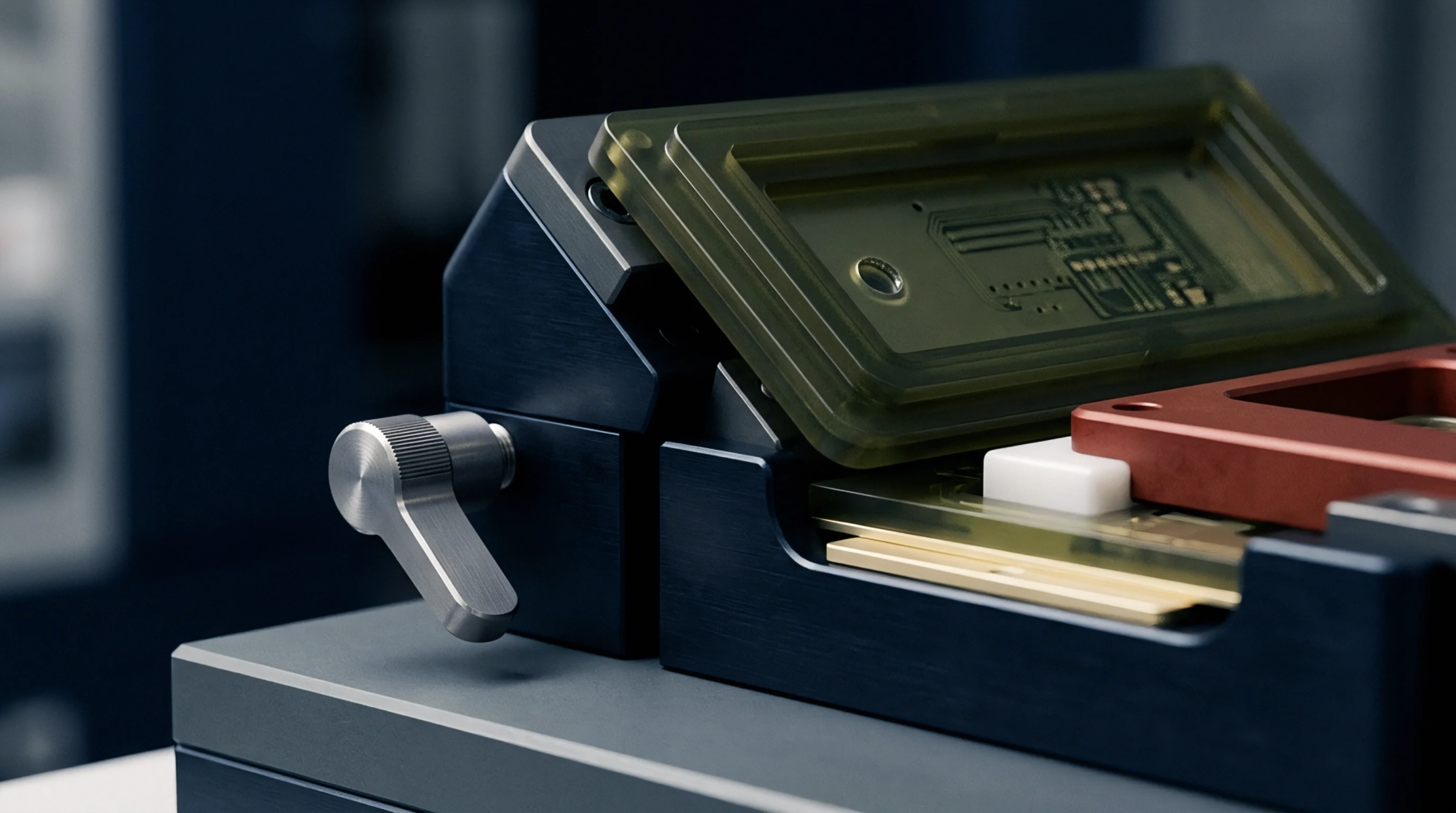
Radiographic Resolution
High energy photons emitted from an extremely small focal spot identify microscopic voids and connectivity anomalies within multilayer circuit boards. A microfocus x-ray system functions by directing a concentrated electron beam onto a metallic target, typically tungsten, to generate a point source of radiation that allows for magnification levels exceeding one thousand times without blurring the edges of the image. This geometry provides the geometric sharpness necessary to resolve features such as ball grid array solder joints and plated through hole barrel integrity where standard industrial imaging lacks the spatial fidelity to isolate individual defects.
Imaging Mechanism
Secondary electrons produced during the beam interaction provide contrast based on the atomic density of the materials inside the housing. The detector array collects transmitted photons while software algorithms reconstruct these intensities into a two dimensional projection or a three dimensional volumetric model through computed tomography. Voids in solder joints appear as darker regions because the material volume through which the beam passes is lower than the surrounding metallic alloy.
Precise motion control stages rotate the sample through several hundred angles to eliminate shadowing effects and separate superimposed circuit features. Resolution limits remain bound by the physical size of the electron beam impact point, as any increase in the source diameter necessitates a reduction in magnification to maintain acceptable signal clarity.
Acceptance Boundary
Quality assurance protocols define the thresholds for defect detection based on the total surface area of porosity or the diameter of inclusions within an interconnection. Acceptable standards allow for minor dispersed voids provided the cumulative total does not reduce the mechanical integrity of the joint or exceed the percentage limits defined in industry assembly specifications. False negatives occur when the orientation of a crack aligns parallel to the beam path, masking the separation from the detector.
Operators calibrate the threshold settings against known reference samples to verify that the equipment maintains the sensitivity required for high density interconnect validation. Constant voltage maintenance in the cathode array ensures the reliability of image acquisition during high volume production cycles.