Spatial Magnification Calibration Fundamentals for Surface Mount X-Ray Inspection
X-ray spatial magnification relies on precise distance ratios, focal spot limits, and grid calibration to ensure valid SMT solder joint measurement.
Scale
Positioning a printed circuit assembly between an X-ray microfocus tube and a flat-panel digital detector establishes the physical cone-beam enlargement of surface mount interconnects. Geometric magnification operates as a direct ratio of distances along the optical centerline: projection distance from the focal spot to the detector divided by distance from the spot to the component feature sets the scale factor. Because of this geometry, slight movements along the primary beam axis produce substantial shifts in dimensional projection.
At high magnification, shifting the stage vertically by fifty micrometers alters feature dimensions on the image plane by several percentage points.
Because X-ray emission diverges and radiographic systems lack refractive focusing elements, geometric projection is the sole mechanism available to enlarge features such as ball grid array joints or micro-passive terminations. Focus-to-object and focus-to-detector distances govern this spatial geometry. Maintaining measurement fidelity across production batches depends on rigid mechanical stability along that axis, as mechanical play in motor drives or lead-screw backlash introduces spatial variance before image processing algorithms run their first calculation.
At a geometric magnification of one hundred times, a ten-micrometer vertical stage positioning drift alters feature scale calculations by one percent.
Focal spot size directly bounds spatial resolution limits and geometric unsharpness. Microfocus tubes use electron beam optics to focus electrons onto a heavy metal target, creating a source spot between one and five micrometers across. Increasing tube wattage introduces more thermal energy at the target, expanding the electron focus and broadening the effective source spot.
Geometric unsharpness scales linearly with focal spot size and magnification, producing penumbral blurring along solder joint boundaries that complicates automated edge detection.
Spatial magnification calibration protocols map the geometric transfer function across the machine’s vertical envelope. Calibrating spatial dimensions at a single height produces flawed physical measurements whenever multi-axis maneuvers change component elevation, leaving automated algorithms with inaccurate scale factors when converting pixel counts into metric dimensions.
| Focal Spot Size (µm) | Magnification Factor | Focus to Object Distance (mm) | Focus to Detector Distance (mm) | Geometric Unsharpness (µm) |
|---|---|---|---|---|
| 1.0 | 10x | 60.0 | 600.0 | 9.0 |
| 1.0 | 50x | 12.0 | 600.0 | 49.0 |
| 3.0 | 50x | 12.0 | 600.0 | 147.0 |
| 3.0 | 100x | 6.0 | 600.0 | 297.0 |
| 5.0 | 100x | 6.0 | 600.0 | 495.0 |
Uncalibrated spatial drift propagates directly into structural pass-fail evaluations. An operator evaluating quad-flat no-lead center ground pads under shifted scale settings risks accepting void levels that exceed allowable limits or rejecting compliant solder fillets.

Grid
Precision calibration targets etched with chromium dot matrix arrays establish the pixel-to-millimeter transformation matrix for digital radioscopy systems. Glass or quartz substrates carry photo-lithographically defined features traceable to international standards. Placed within the chamber, the target allows the system to map spatial scale factors across the active field of view, matching measured pixel spans between feature centers against certified physical distances to generate dimensional mapping coefficients.
Internal thermal dynamics in high-voltage chambers introduce mechanical instability during sustained shifts. Microfocus tubes run hot, radiating heat into the enclosure where aluminum stage supports expand at twenty-three parts per million per degree Celsius. A temperature rise of five degrees Celsius across a four-hundred-millimeter stage frame causes forty-six micrometers of linear expansion, shifting the focus-to-object distance without registering on axis encoders.
- Thermal Expansion Drift drives mechanical movement of stage lead screws, shifting the physical Z-axis location of the board target relative to the source.
- Detector Sensor Tilt causes localized spatial scale distortion across flat panel digital sensor matrices due to non-parallel plane alignment.
- Manipulator Stage Backlash introduces spatial positioning non-repeatability during directional moves along primary axes.
- X-Ray Target Erosion widens the electron spot size over extended operational hours, increasing penumbral blur and altering edge detection thresholds.
Periodic spatial recalibration accounts for this thermal growth and structural settling over time. While flat-panel detectors avoid the severe geometric distortion characteristic of older image intensifiers, mild pin-cushion and barrel distortions still appear toward frame perimeters, requiring polynomial correction grids to maintain uniform scale across every quadrant.
Calibration routines executed at ambient room temperature lose validity as internal cabinet temperatures stabilize during continuous production.
Factory geometric calibration does not eliminate the need for ongoing line calibration. Mechanical settling, floor vibration, and cabinet thermal cycles shift optical scale vectors past acceptable quality thresholds during extended runs, while baseline pixel response requires characterization at actual operating temperatures once target arrays thermally stabilize.

Tilt
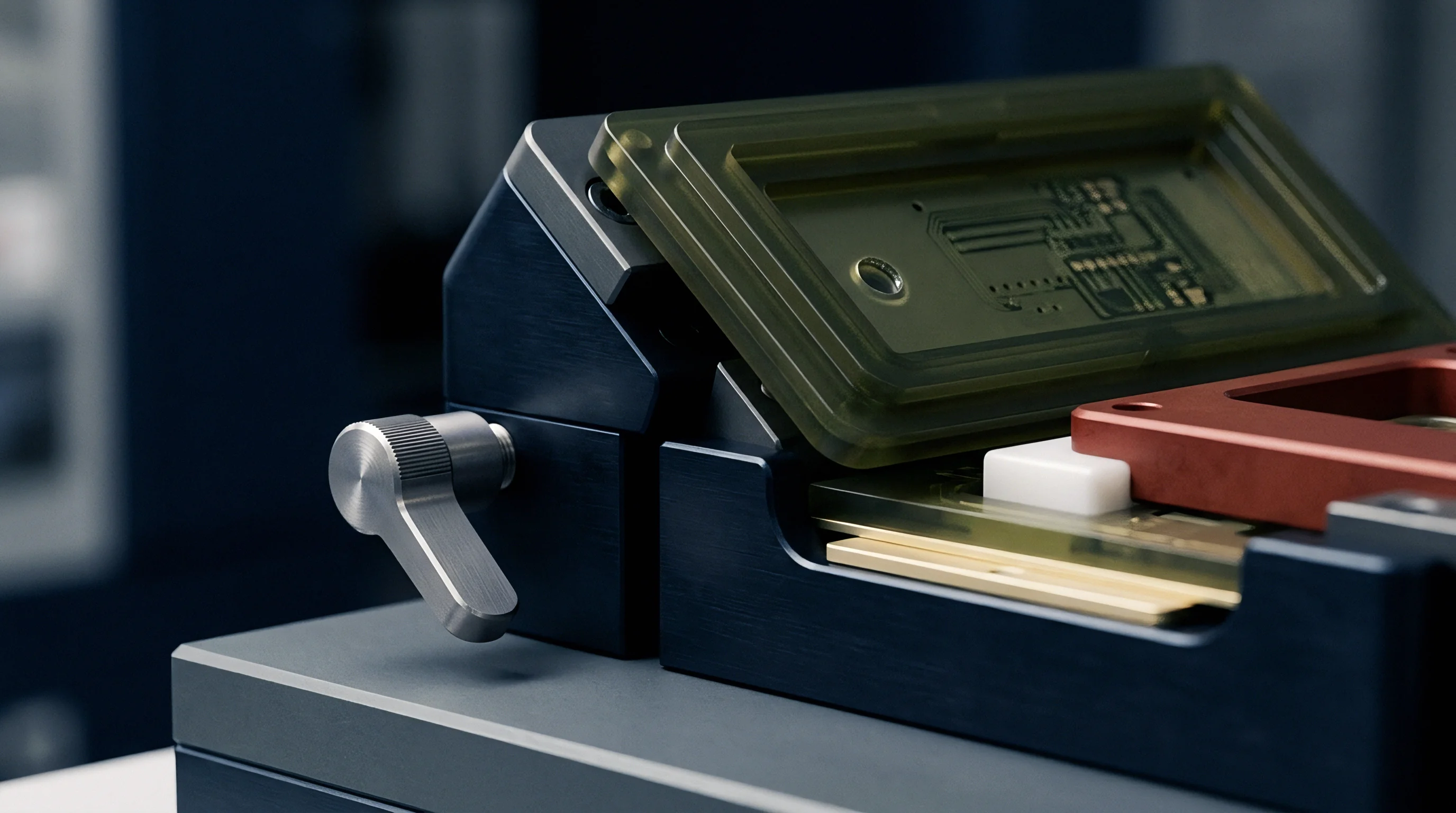
Tilting the board stage or rotating the tube axis introduces non-isometric spatial perspective across the field of view. Oblique viewing exposes structural features unavailable from top-down angles, including heel fillet wetting heights, component tilt, and barrel fill in plated through-holes. However, angling the inspection plane stretches circular solder joints into elliptical silhouettes, and features closer to the source experience higher magnification than those further down the tilt slope.
How Does Oblique Angle Inspection Alter Spatial Magnification Accuracy?
Oblique imaging requires three-dimensional perspective correction because standard planar equations fail as tilt angles steepen. Simple trigonometric scaling ~ dividing measured image lengths by the cosine of the tilt angle ~ presumes an orthogonal, parallel beam path. Microfocus cone-beam radiation diverges continuously, meaning features at the far edges of an angled detector undergo non-linear magnification distortion driven by unequal path lengths from the focal origin.
Multi-axis manipulation stages counter this by applying spatial transformation matrices derived from kinematic modeling. This mirrors calibration methods used on coordinate measuring machines, where laser interferometry corrects non-orthogonal axes and thermal growth vectors. Applying those structural coordinate corrections to cone-beam data preserves scale integrity as stage angles change during automated inspection cycles.
- Position the certified NIST-traceable grid target on the manipulator stage plane at zero degrees tilt.
- Execute a baseline zero-degree spatial calibration to establish the optical reference center point and primary pixel resolution scale.
- Rotate the stage assembly to the target tilt orientation required for production component inspection.
- Capture radiographic image frames across multiple rotational azimuth angles to sample distortion vectors across the field boundaries.
- Compute localized spatial transformation polynomials to update edge tracking algorithms for perspective projection distortion.
Because oblique angles stretch joint contours into ellipses and lengthen ray paths across the detector, standard planar assumptions fail off axis. Without real-time optical displacement tracking, mechanical play during continuous rotational repositioning makes it difficult for automated algorithms to maintain sub-five-micrometer accuracy.
Computation
Converting grayscale attenuation gradients into physical dimensions requires edge-detection routines matched to absolute spatial parameters. In digital radiography, attenuation registers as pixel intensity: dense solder alloys absorb photons and appear dark, whereas fiberglass and resin allow transmission, reading bright. Algorithms locate physical boundaries by calculating grayscale intensity gradient peaks across these transition zones.
Magnification calibration errors compound area calculations quadratically. Assessing voiding beneath bottom-terminated components and ball grid arrays requires dividing total void area by joint contact area, meaning linear scale errors are squared in two-dimensional results. A linear miscalibration of three percent produces a six-point-one percent error in computed void area ratios, skewing assembly compliance evaluations against workmanship standards.
| Package Configuration | True Joint Area (mm²) | True Void Ratio (%) | Uncalibrated Scale Drift | Reported Void Ratio (%) | Class 3 Pass/Fail Result |
|---|---|---|---|---|---|
| BGA Ball (0.40 mm) | 0.126 | 22.0 | +2.0% Scale Shift | 22.9 | Pass |
| BGA Ball (0.40 mm) | 0.126 | 24.0 | +3.5% Scale Shift | 25.7 | False Reject |
| QFN Ground Pad | 16.00 | 28.5 | -3.0% Scale Shift | 26.8 | False Accept |
| QFN Ground Pad | 16.00 | 29.0 | +2.5% Scale Shift | 30.5 | False Reject |
On a BGA pad with a true nominal diameter of four hundred micrometers, an uncalibrated optical zoom setting that scales the matrix by five percent shifts an acceptable twenty-three point five percent void measurement to a failing twenty-five point nine percent. That false reject interrupts production and creates unnecessary rework costs. Negative scale drift produces the opposite failure mode, disguising out-of-spec voiding and releasing non-compliant assemblies into shipment lots.
Contract requirements referencing IPC-A-610 Class 3 specifications invalidate inspection findings derived from spatial systems lacking documented calibration records.
Spatial measurement accuracy depends on stable grayscale threshold values paired with certified geometric scale factors. Running calibration routines on uniform alloy targets establishes repeatable edge-detection thresholds across production batches, preventing threshold shifts from distorting boundary calculations.

Dossier
Calibration records traceable to national metrology standards provide the objective foundation for quality sign-offs in contract electronics manufacturing. Line qualification procedures require documented proof that X-ray dimensional measurements match physical geometry within defined tolerances, giving sourcing auditors the verification needed before approving commercial production.
Maintaining an audit-ready qualification record requires systematic verification of stage mechanics and spatial repeatability across scheduled maintenance intervals, ensuring first-article acceptance and preventing lot rejections during quality reviews.
- Traceable Target Certification documents NIST or equivalent standard reference numbers for physical calibration plates used on the inspection line.
- Spatial Repeatability Logs track variance across twenty consecutive static measurements of target test features to verify mechanical stage precision.
- Thermal Stabilization Times establish required system warm-up durations prior to executing baseline spatial calibration runs.
- Focal Health Indicators monitor spot size changes and beam intensity degradation metrics over operating tube lifespans.
Quality assurance policies mandate that spatial calibration steps execute after any hardware maintenance event, X-ray tube filament replacement, mechanical collision, or software updates. Contract documentation must include explicit calibration clauses defining clear operational bounds for spatial data acceptance.
Standard quality agreements specify that X-ray measurement data holds validity only when captured on equipment calibrated within twelve hours prior to production execution using certified target artifacts.