Quantitative Depth Profiling Procedures for Multi-Layer Circuit Board Surface Finishes
Quantitative depth profiling via AES or XPS accurately measures sub-nanometer interfacial oxidation and phosphorus enrichment to prevent joint failures.

Strata
Surface finishes on multi-layer printed circuit boards use thin engineered films to protect underlying copper pads, allow solder wetting, and enable wire bonding. High-density interconnect designs rely on specific chemical stacks with layer thicknesses ranging from a few nanometers to several micrometers. The main surface finish architectures are Electroless Nickel Immersion Gold, Electroless Nickel Electroless Palladium Immersion Gold, Immersion Silver, Immersion Tin, and Organic Solderability Preservatives.
Measuring total film thickness with bulk analytical methods falls short for material qualification, since mechanical integrity and electrical performance depend directly on subsurface concentration profiles.
Evaluating multi-layer boards requires analytical methods that can profile elemental distribution down through the pad surface. Standard energy-dispersive X-ray fluorescence instruments integrate signal over depths exceeding two micrometers, making them unable to isolate interfacial chemical transitions occurring across five-nanometer boundaries. Quantitative depth profiling resolves atomic distributions across metallic and dielectric interfaces to identify interphase boundaries, oxidation fronts, and impurity segregation bands that govern solder joint reliability.
Metallic Depositions in Multi-Layer Board Fabrication
Coating selection sets the boundary between raw copper lands and assembly materials. Electroless Nickel Immersion Gold deposits a mid-phosphorus or high-phosphorus nickel alloy layer directly onto etched copper pads, followed by a thin displacement layer of pure gold. This nickel deposit acts as a diffusion barrier to stop copper migrating into solder joints.
Phosphorus content within the nickel matrix ranges from 7.0 to 10.0 weight percent, or 11.0 to 15.0 atomic percent. During displacement plating, gold ions oxidize surface nickel atoms, replacing them with metallic gold until a continuous gold film forms and halts the reaction.
Electroless Nickel Electroless Palladium Immersion Gold adds an intermediate palladium layer between the nickel and gold. Palladium prevents galvanic hyper-corrosion of the nickel during gold plating, creating a multi-layer stack suitable for lead-free soldering as well as gold or aluminum wire bonding. Depending on bath formulation chemistry, electroless palladium layers contain co-deposited phosphorus or boron that produce distinct elemental concentration bands.
Immersion silver deposits form homogenous metallic silver coatings with organic sulfur-inhibiting co-additives. Immersion tin produces a dense tin coating over copper, though solid-state reactions between tin and copper form intermetallic phase layers right after plating.
| Finish Type | Layer Architecture | Nominal Layer Thickness | Critical Interfacial Target | Profiling Depth Resolution Target |
|---|---|---|---|---|
| ENIG | Au / Ni-P / Cu | Au: 40-100 nm, Ni-P: 3.0-6.0 µm | Ni-P to Au interface and P concentration spike | < 2.0 nm |
| ENEPIG | Au / Pd-P / Ni-P / Cu | Au: 20-50 nm, Pd-P: 50-150 nm, Ni-P: 3.0-6.0 µm | Pd to Au and Ni-P to Pd transition zones | < 1.5 nm |
| Immersion Silver | Ag + Organic / Cu | Ag: 150-400 nm | Ag surface oxide/sulfide layer and Ag-Cu boundary | < 3.0 nm |
| Immersion Tin | Sn / Cu6Sn5 / Cu | Sn: 800-1200 nm | Sn surface SnO/SnO2 oxides and Cu6Sn5 IMC growth | < 5.0 nm |
| OSP | Organic Azole / Cu | Azole film: 200-500 nm | Carbon-nitrogen breakdown zone and Cu oxide interface | < 2.0 nm |
Elemental Gradient Dynamics across Plated Boundaries
Interfacial chemical composition shifts during assembly thermal cycles. Reflow soldering exposes surface finishes to temperatures exceeding 240 degrees Celsius, driving rapid solid-liquid and solid-solid interdiffusion. In Electroless Nickel Immersion Gold finishes, molten tin-based solder dissolves the thin gold layer within seconds and reacts directly with the nickel-phosphorus deposit to form Cu3Sn4 or Ni3Sn4 intermetallic compounds.
Because phosphorus atoms cannot enter the intermetallic crystal lattice, phosphorus accumulates in an adjacent thin layer.
IPC-4552B specification limits gold thickness to a maximum of 0.10 micrometers to prevent high-gold brittle solder joint formation during SAC307 reflow.
Phosphorus concentrations within this boundary zone build up during thermal processing. Once localized phosphorus exceeds 12.0 atomic percent, the nickel-phosphorus layer shifts from an amorphous structure to crystalline Ni3P. Crystalline nickel phosphide has a high mechanical modulus and breaks brittly.
Quantitative depth profiling tracks the spatial distribution and peak concentration of phosphorus across this intermetallic interface, showing joint degradation risks before micro-cracks form under environmental stress.
Organic Solderability Preservatives rely on single-layer or multi-layer organometallic complexing on copper surfaces. Substituted benzimidazole or imidazole compounds coordinate with copper ions to form protective films. Thermal exposure over repeated reflow cycles decomposes these organic ligands, allowing oxygen to diffuse toward the copper substrate.
Quantitative profiling tracks carbon, nitrogen, copper, and oxygen signal gradients from the surface into the bulk substrate, pinpointing how deep thermal degradation and oxidation have reached.
IPC-4552B Clause 3.2.2 limits maximum allowable immersion gold thickness to 0.10 micrometers, shifting financial liability for hyper-corrosion defects to the plating vendor whenever depth profiles show excess gold deposition.
Sputter
Ion beam etching removes material from circuit board test coupons in a controlled way, exposing subsurface chemical architectures nanometer by nanometer. Inert gas ions generated in an ion source accelerate toward the sample surface under high electrical potentials. Collisions between incident ions and target atoms transfer kinetic energy, driving atomic collision cascades through the top layers of the surface finish.
When the energy transferred to a surface atom exceeds its binding energy, that atom ejects into the vacuum, exposing the layer underneath.
Precision depth profiling requires continuous or sequential ion erosion matched with synchronized spectroscopic measurements. Monoatomic argon ions operating between 0.5 kiloelectronvolts and 4.0 kiloelectronvolts serve as the standard primary beam source for metallic coating profiles. Lower beam energies reduce ion implantation depth and atomic mixing, keeping interfacial chemical gradients sharp.
Argon gas cluster ion beams ~ containing clusters of several hundred to several thousand argon atoms carrying a single positive charge ~ distribute kinetic energy across all constituent atoms, which minimizes surface damage when profiling soft metallic and organic OSP layers.

Ion Beam Energy and Crater Erosion Dynamics
Erosion rates depend on projectile mass, kinetic energy, incident angle, target atomic density, and elemental surface binding energy. Gold has a high sputtering yield under argon ion bombardment, sputtering much faster than nickel or copper under identical beam parameters. Silver comes off quickly, while metal oxides like nickel oxide or tin oxide resist removal.
Because sputtering yields vary across heterogeneous interfaces, converting etching time into depth using a single static erosion rate distorts the depth scale.
Crater morphology dictates analytical depth resolution through deep profiles. Deflecting the focused primary ion beam electronically across a square area produces a flat-bottomed crater. Analytical signals must be collected strictly from the center of this crater floor to exclude secondary electrons or ejected ions coming off sloping sidewalls.
Raster dimensions typically range from 1.0 millimeter by 1.0 millimeter down to 200 micrometers by 200 micrometers. Alignment errors create uneven crater floors, which artificially broadens measured interface widths and hides sharp changes in concentration.
- Primary Ion Beam Alignment ~ Align ion gun optics so the beam stays centered on the analytical aperture of the electron analyzer or optical collector.
- Raster Area Calibration ~ Adjust deflection plate voltages to maintain a uniform raster pattern spanning at least four times the analytical spot size.
- Energy Optimization Sweep ~ Set primary beam energy to 1.0 kiloelectronvolt for gold and palladium layers, dropping to 0.5 kiloelectronvolts near intermetallic interfaces to limit atomic knock-in mixing.
- Electronic Gating Verification ~ Use electronic signal gating to record spectral data only while the ion beam sweeps the central 20 percent of the raster area.
- Crater Depth Profilometry ~ Measure final crater depth using contact stylus profilometry or optical white-light interferometry at the geometric center of the etched floor.
Depth Scale Calibration Using Certified Reference Materials
Converting ion etching duration into physical depth takes careful calibration. Etch rates shift as the sputter front moves from gold into palladium, nickel-phosphorus, and finally the copper substrate. Certified reference materials ~ such as thin tantalum pentoxide films on tantalum substrates or atomic layer deposited multi-layer metallic foils ~ establish baseline sputtering rates under controlled vacuum pressures.
Matrix-specific etch rates must then be calculated for every layer in a complex finish architecture.
Configuring the sputtering beam at two kiloelectronvolts minimizes atomic mixing across the intermetallic boundary. Etch rates vary across phases. Lower beam energies limit atomic knock-in ~ an artifact where incident primary ions push surface atoms deeper into the target substrate.
Knock-in shifts recorded concentration curves deeper into the pad, obscuring true interface locations. Surface roughening from ion bombardment further broadens measured interfaces, while preferential sputtering (where lighter elements like phosphorus etch faster or slower than heavier nickel atoms) alters surface stoichiometry inside the sampling depth.
It remains uncertain whether low-energy argon cluster beams completely eliminate preferential removal of light phosphorus atoms from amorphous nickel matrices during high-resolution interface profiling.


Spectra
Analytical signals collected during surface profiling reveal elemental identity and chemical state transitions through secondary electrons, photoelectrons, optical emissions, or ionized target atoms. Four main depth profiling techniques dominate PCB laboratory characterization: X-ray Photoelectron Spectroscopy, Auger Electron Spectroscopy, Glow Discharge Optical Emission Spectrometry, and Time-of-Flight Secondary Ion Mass Spectrometry. Each technique offers a different mix of spatial, depth, and chemical state sensitivities that suit specific surface finish architectures.
X-ray Photoelectron Spectroscopy identifies chemical oxidation states and elemental ratios by measuring photoelectron kinetic energies under monochromatic X-ray bombardment. Auger Electron Spectroscopy uses a focused electron beam to excite Auger electron emission, delivering spatial resolution down to 10 nanometers. Glow Discharge Optical Emission Spectrometry sputters large sample areas quickly using plasma argon ions, capturing optical spectra from excited target atoms in the plasma.
Time-of-Flight Secondary Ion Mass Spectrometry detects sputtered secondary ions with high-resolution mass spectrometers, reaching parts-per-million detection limits for trace contaminants.

Analytical Modality Comparison for Interfacial Chemical Analysis
Spectroscopic methods differ in spatial footprint and detection thresholds. Auger Electron Spectroscopy offers high spatial resolution, making it possible to profile individual micro-vias, fine-pitch surface mount pads, and micro-crack sidewalls. X-ray Photoelectron Spectroscopy provides moderate lateral resolution but clearly identifies chemical bonding states, distinguishing metallic nickel from nickel oxide and nickel phosphide phases.
Glow Discharge Optical Emission Spectrometry produces rapid depth profiles across five-micrometer ENIG coatings in minutes, which works well for batch quality screening on large test coupons.
Time-of-Flight Secondary Ion Mass Spectrometry offers exceptional surface sensitivity, picking up organic contaminants, azole preserver molecules, and trace halogen ions within the top monolayer of a finish. Because mass spectrometry signal intensity depends heavily on ionization probability, matrix factor adjustments are required to calculate absolute atomic concentrations across changing metal layers.
| Analytical Parameter | XPS / ESCA | Auger (AES) | GDOES | ToF-SIMS |
|---|---|---|---|---|
| Depth Resolution | 1.0 – 3.0 nm | 1.0 – 2.0 nm | 2.0 – 10.0 nm | < 0.5 nm |
| Lateral Analytical Spot | 10 – 100 µm | 10 – 100 nm | 2.0 – 4.0 mm | 50 – 500 nm |
| Chemical State Information | Direct binding energy shifts | Indirect via Auger parameter | None (atomic emission) | Molecular ion fragments |
| Detection Limit | 0.1 – 0.5 at% | 0.1 – 0.5 at% | 1 – 10 ppm | 10 ppb – 1 ppm |
| Analysis Speed (5 µm ENIG) | Slow (2 – 4 hours) | Moderate (1 – 2 hours) | Ultra-fast (1 – 3 mins) | Slow (1 – 3 hours) |
| Light Element Sensitivity (C, O, N, P) | Good | Moderate | Excellent | High (trace levels) |
| Methods note: Capabilities derived under standard argon ion beam profiling conditions at accredited test facilities using monochromatic Al K-alpha XPS sources, 10 kV AES primary electron sources, and liquid metal ion SIMS sources. | ||||

Chemical State Resolution in Intermetallic Compound Profiling
Identifying intermetallic phases requires precise energy spectrum analysis. Photoelectron emission spectra recorded across the nickel-solder interface show binding energy shifts during phase transitions. Metallic nickel displays a primary 2p3/2 photoelectron peak at 852.6 electronvolts.
Nickel oxide shifts to 856.1 electronvolts with prominent shake-up satellite structures. Nickel phosphide compounds formed during reflow show a subtle shift to 853.3 electronvolts.
These chemical shifts make it possible to map oxide scale formation beneath immersion gold layers. In defective ENIG deposits, oxygen profiles show subsurface nickel oxidation that occurred before gold deposition. This internal oxide layer prevents atomic bonding between gold and nickel, causing the gold to peel during thermal stress or wire bonding.
Depth profiling quantifies the integrated area of the oxide peak, offering direct evidence of bath oxidation or improper pre-treatment rinsing.
Matrix Factor Correction and Quantification Accuracy
Converting raw peak intensities into true elemental concentrations requires background subtraction and relative sensitivity factor scaling. High-energy photoelectrons and Auger electrons undergo inelastic scattering while escaping the solid target, creating an elevated spectral background on the low kinetic energy side of photoelectron peaks. Applying Shirley background subtraction isolates true characteristic peak areas from inelastic scattering tails.
- Acquire a baseline energy spectrum across the designated element kinetic energy window.
- Set start and end background energy bounds outside the characteristic peak region.
- Iterate the Shirley background curve calculation until the background matches experimental tails.
- Subtract the background profile from raw spectral data to yield net integrated peak area.
- Apply elemental Relative Sensitivity Factors derived from standard reference compounds.
- Normalize the sum of elemental concentrations to 100 atomic percent for each depth increment.
Nickel-phosphorus layers containing phosphorus concentrations exceeding 11.2 atomic percent undergo catastrophic corrosion when immersion gold plating cycles exceed 14 minutes at 85 degrees Celsius.
Auger depth profiling revealing phosphorus enrichment exceeding 14 atomic percent at the nickel interface signals severe degradation. Signal drift ruins calibration. Quantification algorithms must account for continuous changes in escape depth as sputtering moves from gold into light-element-enriched interface bands.
Ignoring escape depth changes introduces errors up to 25 percent in calculated phosphorus peak values.
Failing to spot secondary ion mass spectrometry instrument drift before testing high-density interconnect substrates leads to severe analytical errors.
Flaw
Structural defects in surface finishes stem from chemical bath imbalances, excessive immersion dwell times, thermal stress, or atmospheric exposure. Defect profiling identifies localized stoichiometry shifts that cause solder joint failures, high contact resistance, or pad de-wetting. Quantitative depth profiling provides the chemical evidence ~ destructive or non-destructive ~ linking observed joint fractures to specific plating anomalies.
Surface defects manifest as hyper-corrosion crevices in electroless nickel deposits, excessive interfacial phosphorus buildup, metallic embrittlement, atmospheric sulfidation, and thermal degradation of organic solderability preserver coatings. Profile signatures reveal specific concentration spikes that serve as forensic proof during supplier liability disputes.

Hyper-Corrosion Crevices and Interfacial Phosphorus Accumulation
Black pad defects in ENIG finishes are among the most severe failure modes in multi-layer circuit board assembly. Hyper-corrosion occurs during displacement gold plating when the immersion gold bath aggressively attacks the underlying electroless nickel. Highly acidic gold baths drive rapid galvanic dissolution of nickel through grain boundaries and microscopic pores in the gold layer, leaving behind an insoluble, highly porous phosphorus-rich network.
Depth profiles recorded through hyper-corroded pads show deep, narrow oxygen and phosphorus spikes extending hundreds of nanometers below the gold-nickel interface. Instead of a sharp step between pure gold and homogenous nickel-phosphorus, the profile exhibits a broad, erratic transition. Gold signals persist deep into the nickel layer alongside elevated phosphorus concentrations exceeding 15 atomic percent and significant oxygen signals.
Depth profiling confirms these hyper-corrosion channels extend completely through the protective nickel layer, compromising pad integrity.
- Black Pad Corrosion Pit ~ Profile shows gold penetration beyond 300 nanometers into nickel, with phosphorus enrichment exceeding 15 atomic percent and co-located oxygen spikes.
- Brittle Phosphorus Layer ~ Profile displays a narrow band of phosphorus exceeding 12 atomic percent directly between the Ni3Sn4 intermetallic layer and the remaining Ni-P bulk.
- Gold Embrittlement Zone ~ Profile reveals residual unreacted gold layers at the intermetallic boundary exceeding 3.0 weight percent total joint solder mass.
- Silver Sulfide Corrosion Film ~ Profile identifies sulfur and silver co-location extending past 50 nanometers into immersion silver deposits, combined with subsurface copper migration.
- Immersion Tin Oxide Scale ~ Profile shows a dual SnO and SnO2 surface oxide layer exceeding 5.0 nanometers in thickness, blocking solder flux wetting.
Thermal Degradation of Preservative and Immersion Coatings
Thermal cycling during multi-pass reflow induces progressive degradation of temporary organic and immersion finishes. Organic Solderability Preservatives rely on protective alkylbenzimidazole molecules complexed onto metallic copper. Exposure to assembly reflow temperatures exceeding 200 degrees Celsius breaks carbon-nitrogen bonds, evaporating volatile organic components and breaking down the thin film over copper topography peaks.
Sputter depth resolution degrades linearly as the ion-etched crater floor develops microscopic surface roughness.
Depth profiling of thermally stressed OSP surfaces reveals marked reductions in carbon and nitrogen profile depths, alongside copper oxidation extending from the outer surface into the pad bulk. When copper oxide thickness beneath degraded OSP exceeds 3.0 nanometers, standard mild rosin fluxes fail to remove surface oxides, leading to severe solder de-wetting and open joints. Similarly, immersion tin coatings undergo solid-state reactions during thermal storage, growing Cu6Sn5 intermetallic layers that consume pure tin.
Depth profiles quantify remaining unreacted pure tin above the intermetallic front, establishing whether enough solderable tin remains for second-side reflow.
Crater rim shadowing occurs whenever beam raster dimensions drop below fifty micrometers. As corrosion extends into nickel and thin films degrade, solderability drops to zero. Sputtering parameters must remain locked across comparative sample runs to prevent instrumental broadening from masking narrow corrosion fissures.
Elevated interfacial phosphorus can stem from sputter-induced selective etching rather than chemical bath displacement anomalies.

Bench
Laboratory execution demands strict physical alignment of test coupons, rigorous vacuum management, and precise secondary ion or electron collection geometries. Raw circuit board panels cannot enter ultra-high vacuum chambers without prior mechanical preparation. Operators extract target pad structures using precision diamond micro-saws, sizing test coupons to fit multi-specimen holders.
Sample cleanliness dictates analytical success in high-resolution depth profiling. Residual drilling dust, handling oils, and volatile organic solvents desorb under high vacuum, depositing thin hydrocarbons onto test coupon surfaces. These hydrocarbon films obscure initial depth profile data points and distort surface concentration calculations for oxygen and carbon.
Test coupons undergo ultrasonic solvent cleaning in high-purity isopropyl alcohol followed by a dry nitrogen blow before entering load locks.
How Do Raster Sizes Distort Interfacial Width Measurements?
Etch crater dimensions dictate whether sputtered sidewall atoms redeposit into the central analytical zone. Small raster areas reduce etching time but introduce severe crater wall effects. When primary ion beams raster over areas smaller than 100 micrometers by 100 micrometers, secondary electrons or photoelectrons generated near crater edges enter the detector collection cone.
Material sputtered from upper gold layers redeposits onto the lower nickel crater floor, creating artificial elemental tails that blend distinct interface steps into gradual slopes.
Expanding raster dimensions to 1.0 millimeter by 1.0 millimeter eliminates sidewall redeposition interference, ensuring measured emissions originate exclusively from a flat, homogenous etching front. Electronic gating restricts detector signal acquisition to the central 10 to 20 percent of the total raster area, cutting off collection while the primary ion beam decelerates or turns at raster boundaries to maintain depth resolution across deep multi-layer profiles.
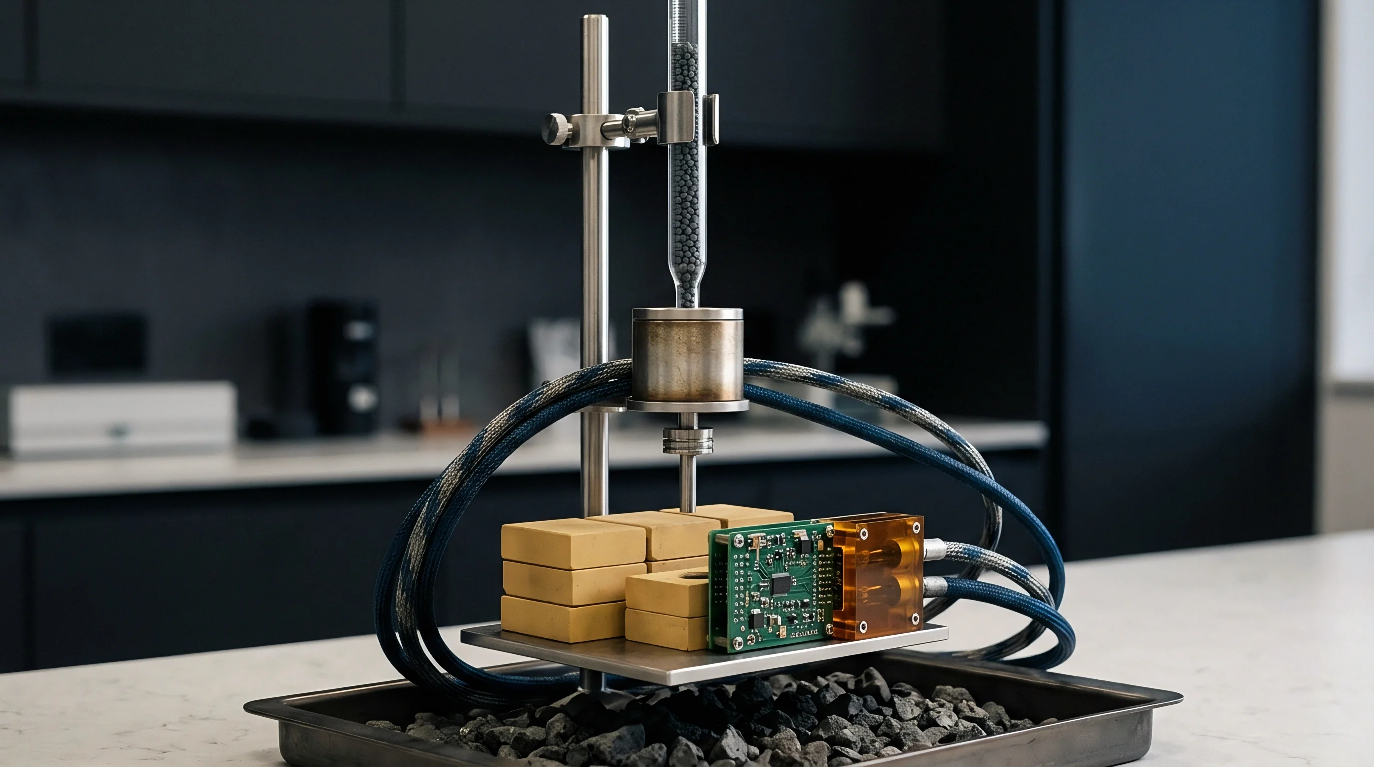
Sample Mounting and Charge Compensation Procedures
Insulating substrate materials require active charge neutralization during electron and ion beam profiling. Circuit board laminates, solder mask dams, and glass-epoxy substrates have high electrical resistivity. Bombarding non-conductive areas with positive ion beams or high-energy electron beams induces localized charge accumulation.
Surface charging deflects incoming primary ion beams and shifts photoelectron energy peaks unpredictably, corrupting quantitative chemical analysis.
- Mount excised test coupons onto metallic sample holders using conductive copper or aluminum vacuum-grade tape.
- Run conductive silver paint bridge lines from the target pad metal frame to the grounded sample holder plate.
- Position low-energy electron flood guns angled directly toward the analytical sampling point.
- Adjust electron flood gun filament current and acceleration voltage to balance incoming ion charges without causing electron beam damage.
- Monitor photoelectron background peak energy alignment continuously to verify stable surface electrostatic potential during sputtering.
Auger electron emission escapes from the upper three nanometers of the target material, offering superior interface localization compared to energy dispersive spectroscopy.
Energy shifts track sample oxidation and rapid gold migration. Maintaining signal stability requires continuous vacuum monitoring below 10 to the power of minus eight millibar. Pressure spikes during ion beam operation indicate gas discharge instability, requiring immediate ion source beam tuning before profile data acquisition continues.
Sharpness at metallic interfaces improves when sputter beam energies are reduced as the etching front approaches phase transitions.
Audit
Verification files accompanying production batches must demonstrate compliance with numerical layer boundaries before shipments receive commercial sign-off. Cross-border electronics procurement practices mandate rigorous proof of delivery, where surface finish compliance reports form an essential part of the technical release dossier. Relying on basic certificate of conformance documents from plating subcontractors exposes buyers to severe field failure risk and financial loss.
Quantitative depth profiling serves as the final arbiter during batch qualification and failure litigation. When non-destructive X-ray fluorescence inspection flags borderline gold thickness or unusual density variations, destructive Auger or XPS depth profiling provides unambiguous atomic composition profiles that validate or reject batch shipments.
Batch Conformity Verification and Specification Guard Bands
Commercial acceptance limits must incorporate guard bands for analytical measurement uncertainty. Standard specifications like IPC-4552B for ENIG and IPC-4556 for ENEPIG define strict upper and lower thickness limits alongside acceptable phosphorus concentration ranges. Laboratory depth profiling measurements carry inherent uncertainty stemming from sputter rate variations, roughness evolution, and signal calibration errors.
Applying guard banding rules reduces acceptance boundaries by the expanded measurement uncertainty value. If a specification dictates a minimum gold thickness of 40 nanometers and instrument measurement uncertainty is calculated at 3.0 nanometers, the operational acceptance threshold rises to 43 nanometers. Guard banding ensures accepted batches fully comply with physical standards, shielding buyers from non-conforming materials that pass only because of favorable measurement tolerances.
| Evaluation Metric | Standard Specification Band | Operational Guard-Banded Limit | Non-Conformity Action | Commercial Financial Allocation |
|---|---|---|---|---|
| ENIG Gold Thickness | 0.04 – 0.10 µm | 0.045 – 0.092 µm | Reject lot / Solderability quarantine | Supplier absorbs re-plating cost |
| ENIG Interfacial P Peak | 7.0 – 10.0 wt% bulk (< 12.0 at% peak) | < 11.2 at% peak at Ni-P/IMC boundary | Immediate batch rejection | Full batch scrap and expedited freight liability |
| ENEPIG Palladium Layer | 0.05 – 0.15 µm | 0.058 – 0.138 µm | Hold lot for wire bond testing | Supplier pays re-testing fees |
| Immersion Silver Sulfide Depth | < 5.0 nm surface Ag2S | < 3.0 nm surface Ag2S | Bake and re-profile or reject | Plating shop pays baking and audit fees |
| Immersion Tin Surface Oxide | < 5.0 nm SnO/SnO2 total | < 3.8 nm total oxide scale | Reject for fine-pitch assembly | Vendor covers production delay penalties |
| Summary note: Commercial thresholds applied across high-reliability automotive and aerospace multi-layer board procurement contracts. Non-conforming test results automatically trigger financial debit notes against supplier quality reserves. | ||||

Commercial Cost Allocation for Destructive Depth Profiling
Analytical depth profiling carries substantial laboratory costs that must be factored into manufacturing budgets. Standard high-resolution AES or XPS depth profiling routines cost between 800 and 1500 US dollars per test pad location, reflecting instrument capital costs and specialized operator hours. Conducting routine depth profiling on every production panel is financially unviable for standard commercial assemblies.
Cost-effective quality assurance mandates a layered testing strategy. Non-destructive micro-XRF screens 100 percent of production lot test coupons to track high-level layer thickness trends. Destructive quantitative depth profiling is reserved for initial process qualification, chemical bath changeovers, high-risk aerospace lot releases, and forensic auditing of lots flagged during XRF screening.
Once initial deposits pass audit, suppliers face strict depth profiling limits before full production releases receive sign-off. This hybrid quality verification model balances analytical costs against product failure exposure, ensuring complete technical compliance while keeping production costs manageable.
Commercial contracts incorporating quantitative depth profiling criteria eliminate ambiguities in failure analysis, ensuring chemical degradation at metallic interfaces is assigned directly to bath chemistry control or improper thermal processing.